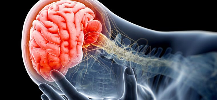
It’s been called the “X-ray of the 21st century” because it does for the brain what X-rays do for bones.
A new type of brain scan being researched at the University of Pittsburgh, displayed Thursday morning at Pitt-Johnstown, gives doctors a detailed look at what one might call the “wiring” of the brain, making it much easier to diagnose and understand traumatic brain injuries.
This information could be revolutionary for veterans who return home with traumatic brain injuries, because doctors could help them better understand the effects of what are sometimes called “invisible wounds.”
“This was the first opportunity to actually see inside the brain to see how injured a veteran was,”
said state Rep. Bryan Barbin, a Democrat who represents Cambria and Somerset counties.
Chief scientist Walt Schneider said this scanning technology will not only help veterans see what their injury looks like, but also predict what the effects of the injury might be.
For example, a doctor could tell a veteran’s spouse that the veteran has an injury on a specific part of the brain, like the part that controls anger or emotion.
“We have on person who says it saved their marriage,”
Schneider said. “Because they were warned that there are going to be emotional outbreaks.”
Barbin sponsored a bill in the Pennsylvania House of Representatives, hoping to provide as much as a million dollars for Pennsylvania veterans to participate in research studies of this technology.
“There’s a possibility of showing a spouse, or parents, that there’s this injury that’s real,”
Barbin said. “Just as real as if you lost an arm.”
Barbin also mentioned, though, that that funding is likely on hold for some time as the state continues to grapple over a dramatically under-funded budget.
This technology is still in the research phase, but doctors hope at some point it could also be used for athletes who have concussions.


